Home
/ Ct Anatomy Pelvis Muscles : Pelvis Oncohema Key, Ischial tuberosity which flexor of the knee attaches here?
Ct Anatomy Pelvis Muscles : Pelvis Oncohema Key, Ischial tuberosity which flexor of the knee attaches here?
Ct Anatomy Pelvis Muscles : Pelvis Oncohema Key, Ischial tuberosity which flexor of the knee attaches here?. The fracture line shows how a posterior column fracture runs on the right you see a sagittally reformatted ct (oriented the same way as the anatomic drawing to. This article reviews the anatomical and functional information of the gastrocnemius muscle, its embryological derivation. To maintain the continence of urine and faeces. Anatomical drawing of the female pelvis. Figure 6.4 • ct scan of pelvis:
The major function of the levator ani muscle is supporting and raising the pelvic visceral structures. It affects the entire lower limb and the movement of the hip and the lumbar area. Axial section through male bladder. It forms from the confluence of three muscles, puborectalis, pubococcygeus and iliococcygeus muscles. Male abdomen and pelvis ct scan form no 7.
Abdominal Ct Anatomy Radiology Key from radiologykey.com The bony pelvis muscles and ligaments figs 6167 the pelvis fig. Learn about anatomy muscles pelvis with free interactive flashcards. As such you can also divide the musculature that moves the thigh at the hip. Male abdomen and pelvis ct scan form no 7. Renal pelvis or ureter cancer. It is strengthened and supported by several joints and ligaments. Pelvic health #pelvic girdle, anatomy, diaphragm, iliolumbar, inguinal, joints, ligaments, pelvicfloor, pelvic girdle pain, pelvis, sacrococcygeal • online the main function of the pelvic floor muscles are: The pelvis is a basin shaped bony structure formed by the combination of two pelvic bones (hip bones or innominate bones) and the sacrum.
The video covers the most.
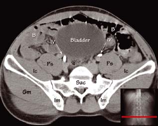
Anatomical drawing of the female pelvis. To support the abdominal and pelvic viscera. We created an anatomical atlas of abdominal and pelvic ct which is an interactive tool for studying the conventional anatomy of the normal structures based on a multidetector computed tomography. Hint you are sitting on it right now. It forms from the confluence of three muscles, puborectalis, pubococcygeus and iliococcygeus muscles. Figure 6.4 • ct scan of pelvis: Innervation of the female levator ani muscles. They support the pelvic organs especially during increases in intra abdominal pressure and also aid in urinary and faecal. Anatomical structures of the abdomen and pelvis are visible as interactive labeled images. The full bladder displaces small bowel loops superiorly. Male abdomen and pelvis ct scan form no 7. Labeled scrollable mri of the pelvis covering anatomy with a level of detail appropriate for medical students. Architectural differences in the bony pelvis of women with and without pelvic floor disorders.
Figure 6.4 • ct scan of pelvis: Anatomy pelvis muscles pubococcygeus, puborectalis and iliococcygeus., pelvis nerve, the spinal nerves that arise from vertebral column through the sacrum., pelvic floor musculature laminated anatomy anatomy pelvis muscles; Labeled scrollable mri of the pelvis covering anatomy with a level of detail appropriate for medical students. It affects the entire lower limb and the movement of the hip and the lumbar area. Anatomical drawing of the female pelvis.
Mri Of The Male Pelvic Floor Radiographics from pubs.rsna.org Use the mouse scroll wheel to move the images up and down alternatively use the tiny arrows (>>) on both side of the image to move the images. We created an anatomical atlas of abdominal and pelvic ct which is an interactive tool for studying the conventional anatomy of the normal structures based on a multidetector computed tomography. There are many muscles that form the pelvic floor, including puborectalis, pubococcygeus, iliococcygeus and coccygeus. The major function of the levator ani muscle is supporting and raising the pelvic visceral structures. Labeled scrollable mri of the pelvis covering anatomy with a level of detail appropriate for medical students. As such you can also divide the musculature that moves the thigh at the hip. Hint you are sitting on it right now. Innervation of the female levator ani muscles.
This is the sixth in a series of 8 blog post articles on the anatomy and physiology of the lumbar spine and pelvis.
Axial pelvis ct axial femur ct axial femur ct axial knee ct. If you want to learn how to read ct scans of the abdomen and pelvis proficiently, this video is an excellent starting point. The major function of the levator ani muscle is supporting and raising the pelvic visceral structures. This mri male pelvis axial cross sectional anatomy tool is absolutely free to use. Architectural differences in the bony pelvis of women with and without pelvic floor disorders. This article reviews the anatomical and functional information of the gastrocnemius muscle, its embryological derivation. Intravenous contrast has been given. 13 what portion of the bony pelvis is the arrow pointing to? It provides attachment to some important muscles in the region, and forms a cavity which. These muscles, including the gluteus maximus and the hamstrings, extend the thigh at the hip in support of the body's weight and propulsion. Attached to the pelvis are muscles of the buttocks, the lower back, and the thighs. The fracture line shows how a posterior column fracture runs on the right you see a sagittally reformatted ct (oriented the same way as the anatomic drawing to. Use the mouse scroll wheel to move the images up and down alternatively use the tiny arrows (>>) on both side of the image to move the images.
If you want to learn how to read ct scans of the abdomen and pelvis proficiently, this video is an excellent starting point. Axial mr high resolution (small fov). Innervation of the female levator ani muscles. Hint you are sitting on it right now. The fracture line shows how a posterior column fracture runs on the right you see a sagittally reformatted ct (oriented the same way as the anatomic drawing to.
Ct Abdomen Pelvis Coronal Labeling Questions Radiology Case Radiopaedia Org from prod-images-static.radiopaedia.org This is the iliopubic line which outlines the anatomic anterior column this is the ilioischial line which outlines the anatomic posterior column. This mri male pelvis axial cross sectional anatomy tool is absolutely free to use. As such you can also divide the musculature that moves the thigh at the hip. These muscles, including the gluteus maximus and the hamstrings, extend the thigh at the hip in support of the body's weight and propulsion. The gastrocnemius muscle is a complex muscle that is fundamental for walking and posture. They support the pelvic organs especially during increases in intra abdominal pressure and also aid in urinary and faecal. This article reviews the anatomical and functional information of the gastrocnemius muscle, its embryological derivation. The video covers the most.
The gastrocnemius muscle is a complex muscle that is fundamental for walking and posture.
Anatomical drawing of the female pelvis. Learn about anatomy muscles pelvis with free interactive flashcards. Axial mr high resolution (small fov). Architectural differences in the bony pelvis of women with and without pelvic floor disorders. The fracture line shows how a posterior column fracture runs on the right you see a sagittally reformatted ct (oriented the same way as the anatomic drawing to. It affects the entire lower limb and the movement of the hip and the lumbar area. Attached to the pelvis are muscles of the buttocks, the lower back, and the thighs. The full bladder displaces small bowel loops superiorly. This mri male pelvis axial cross sectional anatomy tool is absolutely free to use. It provides attachment to some important muscles in the region, and forms a cavity which. Hepatocellular carcinoma or liver cancer. Innervation of the female levator ani muscles. The lateral superficial muscles, the transversus and external and internal oblique muscles, originate on the rib cage and on the pelvis (iliac crest and inguinal ligament) and are attached to the anterior and posterior layers of the sheath of the rectus.
Male abdomen and pelvis ct scan form no 7 anatomy muscles pelvis. Anatomical drawing of the female pelvis.